2. What Is A Cell? What is Cell Research?
Cells are defined as the smallest functional biological unit of all organisms, capable of autonomous replication and dynamic interaction with changing environments. They may exist as forms of independent life, as in the case of bacteria and other unicellular organisms, or be a subunit of multicellular organisms contributing to specialized functions that allow the organism to perform as a whole (HeLa Primer PPT Slides 12-19: History of Cell Biology).
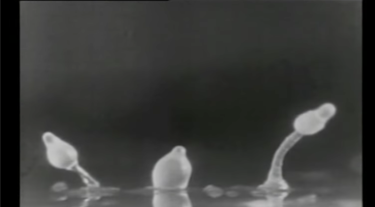
A commonality to all cells is the chemical composition of carbon, nitrogen, oxygen, hydrogen, phosphorous and sulphur. These chemical units can organize to form water, among other compounds, which makes up 70-90% of the total cell volume. These chemical units are also organized into larger molecular polymers that have specific forms and functions, called macromolecules.
There are four types of macromolecules (lipids, nucleic acids, proteins, and sugars).
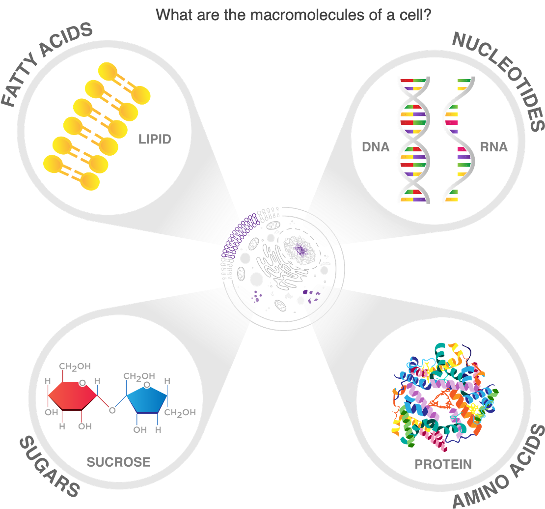
They collectively make up all the components of a cell and allow cells to interact and respond to their environment (HeLa Primer PPT Slide 20: Macromolecules). Fat macromolecules, or lipids, form membranes that create a barrier with the outside environment and form specialized compartments inside the cell. One such compartment is the nucleus, which houses macromolecules called nucleic acids, otherwise known as DNA and RNA. DNA is the genetic material that is passed on from generation to generation, and serves as an information depot for the synthesis of other macromolecules called proteins. Proteins can be subdivided into categories based on function (HeLa Primer PPT Slide 21: Proteins).
Some proteins provide a structural role to the cell that is akin to our skeletons, giving the cell membrane integrity and strength, and are collectively termed cytoskeletal proteins (cyto- meaning cell). Other proteins are involved in transporting material to and from various compartments within the cell and to the outside environment. Still other proteins “communicate” with the outside environment in the form of signaling pathways. A signaling pathway involves many proteins that interact in a specific sequence, in response to an initial signal outside of the cell (extracellular environment). Often this pathway includes a receptor protein that straddles the cell membrane, allowing it to undergo conformational changes upon binding this external signal. This change in conformational shape allows the receptor protein to make new interactions with molecules and proteins inside of the cell. This cascade of interaction elicits a change in cell behavior by triggering protein synthesis, protein activation, and/or protein reorganization resulting in cellular shape changes required for cell division, cell death, cell movement, or cell specialization. To synthesize proteins, a cell responds to an external signal and uses the information in its DNA to build the appropriate proteins for that specific environment. To accomplish this, the cell uses energy, which is created by a collection of proteins called enzymes that break down another macromolecule type, called sugars, to generate molecular energy in the form of Adenosine Triphosphate (ATP).
Cell biology is the study of cells as single units and also as organized collectives, referred to as tissues and organs.
One of the most important aspects of cell biology is the role of the extracellular matrix (ECM), which is the material that surrounds cells in multicellular organisms. The ECM is a scaffold of structural proteins and sugar complexes that serves as a depot for extracellular signals such as growth factors. Cell behavior, structure, and organization are influenced by the composition and structure of the ECM, which varies among different tissues and organs. The ECM structure and composition can also be influenced by aging, trauma, and injury, all of which may halt or stimulate cell regeneration and specialization.
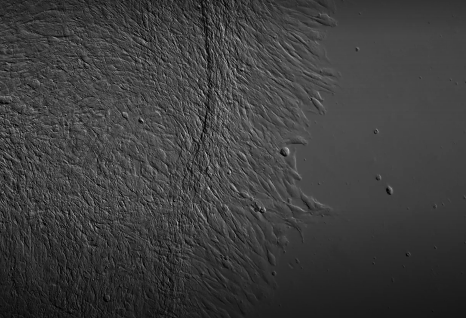
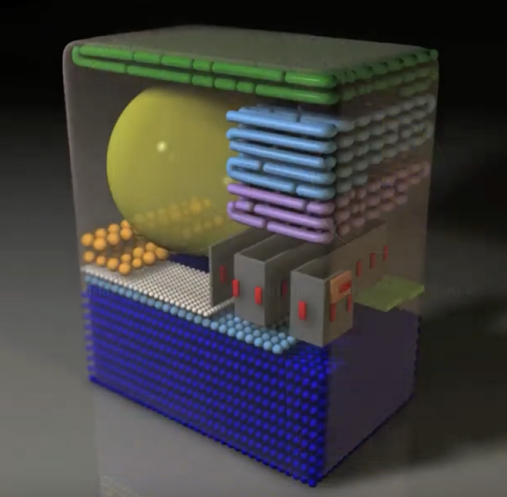
This specialization can lead to dramatically different cell structures, internal compositions, and organizations that retain the ability to respond to the environment dynamically and quickly. The 2011 International Science and Engineering Visualization Challenge People’s Choice 1st Prize in Video “Rapid Visual Inventory & Comparison of Complex 3D Structures” by Graham Johnson et al., illustrates how mouse pancreatic cells rapidly respond to sugar exposure by altering the synthesis and relative distribution of various organelles, which act as specialized sub structures within cells (HeLa Primer PPT Slide 22: Scale and Dynamics).
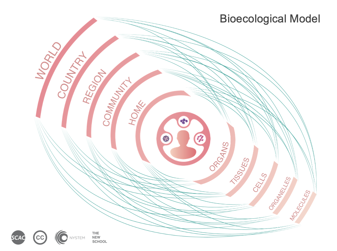
In the human body, there are approximately 200 types of specialized human cells constituting the 10 trillion cells of our body and trillions more bacterial cells and viruses. Thus, the human body is an ecosystem situated within a larger ecosystem that includes our built and social environments, sometimes referred to as the bio- ecological model. Russian-born American psychologist Urie Bronfenbrenner, in his book The Ecology of Human Development: Experiments by Nature and Design published in 1979, first proposed this model (Bronfenbrenner,1979). (HeLa Primer PPT Slide 23/Slide 3: Bioecological Model)
As early as the 1600s, scientists studied cells in living tissues and in isolation from their natural ecosystems, often employing microscopy and visualization techniques to label or dye particular compartments, or organelles, in the cell. (HeLa Primer PPT Slide 24 & 25: How Do We Visualize Cells). As technological and scientific advances were made, cells were not only studied in isolation or in pure liquid culture, but in complex three-dimensional environments that mimicked their natural environments (HeLa Primer PPT Slide 26: Moving from 2D to 3D). One of the most essential technological breakthroughs for biomedical research was the ability to propagate human cells indefinitely in the laboratory, otherwise known as cell culture.
For a deeper history on cell culture we can turn to Boyce Rensberger’s excellent trade book Life Itself: Exploring the Realm of the Living Cell (Rensberger, 1996), Jane Maienschein’s book Whose View of Life? Embryos, Cloning and Stem Cells (Maienschein, 2005), and her related multimedia learning resource The Embryo Project. In addition, Willy Lensch and Christine Mummary provide a review of cell culture techniques as they relate to contemporary stem cell research and, like Maienschein and the NIH report “Regenerative Medicine,” evoke the Greek legend of Prometheus to highlight the ethical controversies associated with “culturing life” (Department of Health and Human Services, 2006; Lensch & Mummery, 2013). Hannah Landecker extends this analysis in her anthropological view of the evolution of cell culture research in Culturing Life: How Cells Became Technologies (Landecker, 2010).
Created : July 23, 2026